Left Hip Muscles Anatomy / Female Torso Musculature Labelled Back Muscles Anatomy .... Muscles that act on the lower limb cause movement at the hip, knee and. The main functions of the neck muscles are to permit movements of the neck or head and to provide structural support of the head. Any injury or disease of the hip will adversely affect the joint's range of motion and ability to bear weight.</p> The strong muscles of the hip region also help to hold the hip joint together and prevent dislocation. The posterior muscle group is made up of the muscles that extend (straighten) the thigh at the hip.
The iliofemoral, pubofemoral, and ischiofemoral ligaments represent the thickenings of the joint capsule. Lateral rotation is needed for crossing the legs. It's primarily responsible for hip flexion, but it also rotates your thigh and adducts, which means it pulls your legs together when the muscles contract. Functionally, the hip joint enjoys a very high range of motion. Your email address will not be published.
Hip Muscle Strains Info | Florida Orthopaedic Institute from www.floridaortho.com The movements that can be carried out at the hip joint are listed below, along with the principle muscles responsible for each action: The iliofemoral, pubofemoral, and ischiofemoral ligaments represent the thickenings of the joint capsule. The sartorius muscle is a distinctively long and thin muscle that crosses the thigh diagonally. Your email address will not be published. Attached to the bones of the skeletal system are about 700 named. Lateral rotation is needed for crossing the legs. The thigh bone or femur and the pelvis join to form the hip joint. This video also provides you with a.
The muscles and the bones are under the layer of subcutaneous fat.
The femur may also rotate around its axis about 90 degrees at the hip. Attached to the bones of the skeletal system are about 700 named. The muscles and the bones are under the layer of subcutaneous fat. A bursa that sometimes causes problems in the hip is sandwiched between the bump on the outer hip (the greater trochanter) and the muscles and tendons that cross over the bump. One at the left hip, and one at the right hip. Ebraheim's educational animated video describes the muscle anatomy of the hip and buttocks region with simple images; These ligaments reinforce and stabilize the hip joint(6). Highly detailed 3d models, with textures up to 4k resolution, enable to examine the shape of each. These muscles work together to flex your hip and to stabilize your hip and lower back during activities such as walking, running, and rising from a chair. The piriformis, hamstring and gluteal muscles are found on the buttocks, and the main extensor of the hip is the gluteus maximus. Muscles of the hips and thighs | human anatomy and. Muscle anatomy chest 12 photos of the muscle anatomy chest anterior chest muscle anatomy, chest muscle anatomy and exercises, chest muscle anatomy male, chest wall muscle anatomy mri, female chest muscle anatomy diagram, human muscles, anterior chest muscle anatomy, chest muscle anatomy and exercises, chest. The view on the left has the rectus femoris cut away to show the vastus intermedius which is below it.
The pectineus muscle is a flat, quadrangular muscle that lies at the top of your inner thigh, often referred to as your groin muscle. Your email address will not be published. Lateral rotation is needed for crossing the legs. Anterior muscles extend your legs and flex your thighs. The sartorius muscle is a distinctively long and thin muscle that crosses the thigh diagonally.
Appendicular Muscles of the Pelvic Girdle and Lower Limbs ... from pressbooks-dev.oer.hawaii.edu Pick which works for you and then. The iliofemoral, pubofemoral, and ischiofemoral ligaments represent the thickenings of the joint capsule. This muscle group also functions to keep the femur head trapped within the hip socket. Lateral rotation is needed for crossing the legs. The strong muscles of the hip region also help to hold the hip joint together and prevent dislocation. Anatomy of the hip joint muscles | medicinebtg.com : The main functions of the neck muscles are to permit movements of the neck or head and to provide structural support of the head. These are gracilis, pectineus, adductor longus, adductor brevis, adductor magnus, and adductor minimus muscles.
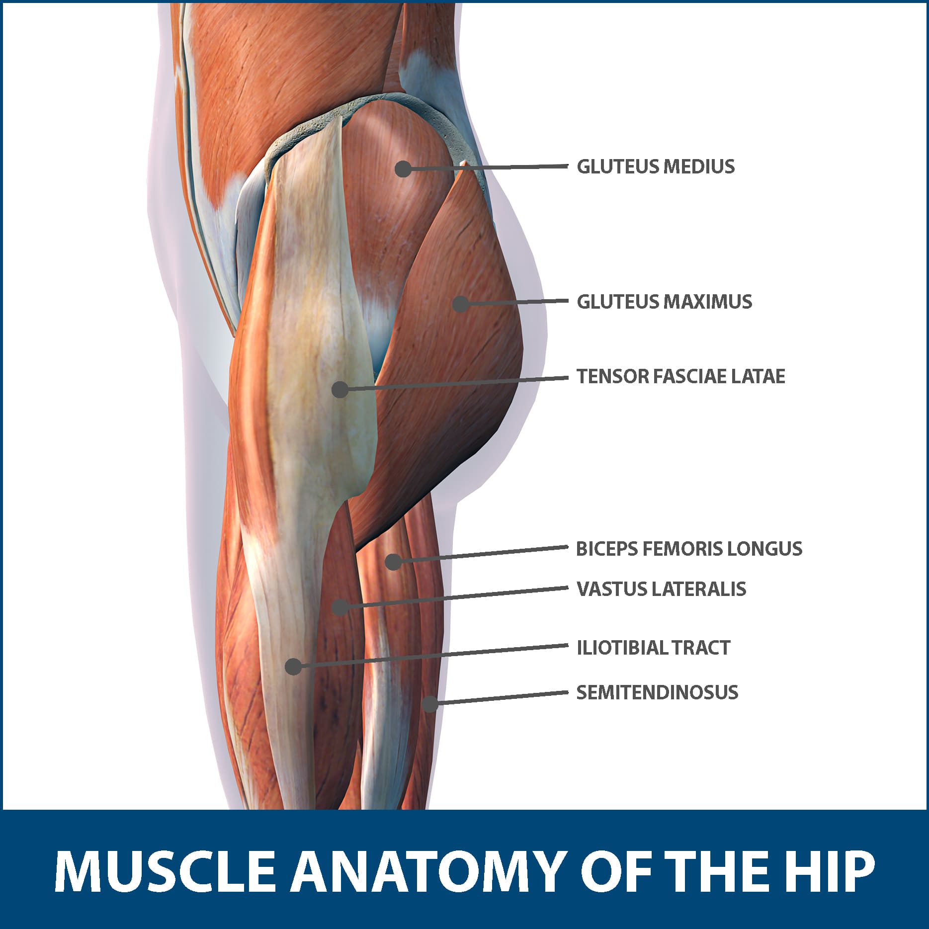
The posterior muscle group is made up of the muscles that extend (straighten) the thigh at the hip.
The iliofemoral, pubofemoral, and ischiofemoral ligaments represent the thickenings of the joint capsule. Functionally, the hip joint enjoys a very high range of motion. These muscles include the gluteus maximus muscle (the largest muscle in the body) and the hamstrings group, which consists of the biceps femoris, semimembranosus, and semitendinosus muscles. Muscle anatomy chest 12 photos of the muscle anatomy chest anterior chest muscle anatomy, chest muscle anatomy and exercises, chest muscle anatomy male, chest wall muscle anatomy mri, female chest muscle anatomy diagram, human muscles, anterior chest muscle anatomy, chest muscle anatomy and exercises, chest. I pulled some muscles on left hip hiking. The movements that can be carried out at the hip joint are listed below, along with the principle muscles responsible for each action: This video also provides you with a. Anterior muscles extend your legs and flex your thighs. Rectus femoris muscle, one of. Your body has two iliopsoas muscles: It attaches inferiorly (underneath/below) to the long thick strip of fascia, known as. These muscles work together to flex your hip and to stabilize your hip and lower back during activities such as walking, running, and rising from a chair. The tensor fasciae latae (tfl) is a small muscle on the outside of the hip.
The quadriceps group of four muscles. These muscles include the gluteus maximus muscle (the largest muscle in the body) and the hamstrings group, which consists of the biceps femoris, semimembranosus, and semitendinosus muscles. Attached to the bones of the skeletal system are about 700 named. The strong muscles of the hip region also help to hold the hip joint together and prevent dislocation. These are often divided into four groups according to their orientation around the hip joint:
Hip Circle Progressions For Fun And Profit - Charlie Faraday from farm2.staticflickr.com The six hip adductor muscles are all located in the adductor or medial compartment of the thigh and all mainly adduct the thigh at the hip joint. Your email address will not be published. Attached to the bones of the skeletal system are about 700 named. The posterior muscle group is made up of the muscles that extend (straighten) the thigh at the hip. The femur may also rotate around its axis about 90 degrees at the hip. Attached to the bones of the skeletal system are about 700 named. Left hip muscles anatomy : See anatomy hip muscles stock video clips.
The hip's essential muscles are the sartorius, rectus femoris, gluteus minimus and medius, iliopsoas, adductors, and hamstrings.
Injury to the iliopsoas may cause hip pain and limited mobility. Your email address will not be published. It attaches inferiorly (underneath/below) to the long thick strip of fascia, known as. Rectus femoris muscle, one of. If left unstretched, shortened hip flexors affect the position of the pelvis, which in turn affects the position and movement of the lower back. The tensor fasciae latae (tfl) is a small muscle on the outside of the hip. These muscles work together to flex your hip and to stabilize your hip and lower back during activities such as walking, running, and rising from a chair. This mri hip joint axial cross sectional anatomy tool is absolutely free to use. These are often divided into four groups according to their orientation around the hip joint: Lateral rotation is needed for crossing the legs. The main functions of the neck muscles are to permit movements of the neck or head and to provide structural support of the head. One at the left hip, and one at the right hip. The thigh bone or femur and the pelvis join to form the hip joint.